
Summarized by : Dr Hani Alhebshi .
Three treatment options for replacing missing lateral incisors :
1.Canine substitution. ( Space Closure ).
2.A tooth supported restoration ( Space Opening ).
3.A single tooth implant ( Space Opening ).
The primary consideration to choose one option is to choose the most conservative one or the least invasive according to each case.
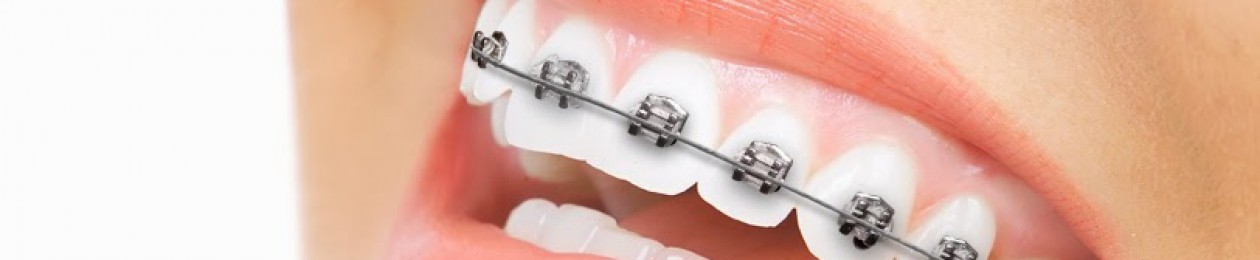
Canine substitution :
Select the appropriate patient for canine substitution according to several criteria :
A. Malocclusion :
Two types of malocclusion can accept canine substitution according to
Kokich:
- Class II with no crowding in mandibular arch.In this pattern molars remain in class II and premolars are located in traditional canine position.
- Class I with crowding in lower arch that necessitate extraction.
Diagnostic wax up will greatly help the orthodontist and the dentist to evaluate the final occlusion and how much canine reduction is necessary.
B.Profile :
- The ideal profile to accept canine substitution is straight profile or mildly convex.
- Moderately convex or retrusive mandible or chin are not appropriate patients.
C. Canine shape & color :
- Canine is much wider and convex labial surface that necessitate significant reduction labially and lingually.
- Restoration of the mesioincisal and distoincisal edges to recreate lateral shape.
- Canine has more saturated color than lateral incisor so it can be changed by bleaching which is the most conservative way. If it fails a veneer
may be indicated. - Finally , a canine with narrow mesiodistal width at CEJ produces a more esthetic emergence profile than if it was wider. So the ideal lateral substitute is a canine that has same color of central incisor , narrow at CEJ buccolingually and mesiodistally , has a relatively flat labial surface and narrow mid crown width buccolingually.
D. Lip level :
- If patient has high lip line , that will increase the demand for correct levelling of the gingival margin.
- The gingival margin of substituted canine has to be placed incisally by extrusion.
- Gingival margin of upper premolars usually are placed more coronally than the central incisor , if this is a concern to the patient , crown lengthening can be performed.
- Patients with high smile lines , canine root eminence could also be an esthetic concern.
E. Treatment and final position:
- Orthodontist should place brackets according to gingival margin height rather than incisal edge or cusp tip. Typically they are placed in a position that will erupt the teeth into the appropriate lateral position.
- As they erupt , premature contacts are reduced periodically untill they reach a suitable position. Bleeching , composite or porcelain veneer can be used to achieve better esthetics.
*******************************************************
2nd option is tooth supported restorations :
Three primary types of tooth supported restorations Are :
- Resin bonded fixed partial denture
- Cantilevered fixed partial denture.
- Full coverage fixed partial denture.
Some patients lack sufficient space for lateral restoration due to ectopic eruption of canine into lateral position. Orthodontist must move the canine to its normal position. This will aid in achieving alveolar ridge development at lateral position.
Determination of appropriate spacing :
Four ways to determine the appropriate space needed for the missing laterals :
- The golden proportion (not accurate method) :
- The perceived width of anterior teeth as viewed from anterior view have a ratio of 1 : 0.62 with the tooth distal to it. For example , photograph of central of 8 mm visually have a lateral incisor of 5 mm ( 0.62 × 8 ) visually too..
- To use the contralateral incisor : if it is present and in good size and shape.
- Bolton Analysis :
- The approximate ratio of lower to maxillary anterior teeth is 0.78.
- By mathematical calculations we can get the supposed lateral incisor width.
- The approximate ratio of lower to maxillary anterior teeth is 0.78.
- Diagnostic wax up :
The most predictable guide for determining ideal spacing is to construct a diagnostic wax up.
Tooth supported restorations :
A. Resin Bonded fixed partial denture :
- The most conservative option.
- This will leave the adjacent teeth relatively untouched.
- Relies solely on adhesion without pins or grooves.
- Success rate varies 45% failure rate over 11 months.
- 10 % failure over 11 years.
- Debonding is the most common cause of failure.
Criteria to consider :
- Position :
- Resin bonded in deep bite cases has high incidence of failure because of high lateral forces.
- Object loaded with shear forces can withstand 40% more load prior to failure as compared to same object loaded with tensile force.
- Mobility of the Abutments :
- It is contraindication in resin bonded due to the stress placed on the bond interface.
- Thickness and translucency of the abutment teeth :
- When retainer extensions are carried too coronal , thin teeth or teeth with high degree of translucency in incisal one third can appear grey due to the show through of the metal retainer.
- Occlusal parafunction :
- Any signs of parafunction contraindicate the use of resin bonded dentures.
B : Cantilevered fixed partial denture :
- Canine is an ideal abutment for cantilever.
- Doesn’t depend on amount of proclination or mobility.
- Retention and resistance requires the use of pins which can affect the pulp especially if pulp size is large.
- Thickness and translucency of the retainer should be evaluated like resin bonded fixed denture.
- The key to success is to remove all contacts in excursive movements on potic . if not removed ; that would cause potential risks of loosening of restorations , migration of abutment and fracture.
C: Convential Full-Coverage fixed partial denture :
- The least conservative option.
- The treatment of choice when replacing an existing fixed denture or when adjacent teeth requires restorations for structural reasons like caries or fracture or to alter the facial esthetics.
Some important points to consider :
- The alignment of the anticipated abutments should be parallel ( canine and central incisors long axis ). If angulations are not parallel , the dentist will have to over prepare the teeth.
- They should be parallel also when viewed from lateral perspective for same reason.
- Orthodontist can help increase the size of the joint by leaving an anterior bite or excess horizontal overjet of approximately 0.5 mm to 0.75 mm.
*******************************************************
3rd Option is Single-tooth Implants :
Diagnosis of missing lateral is commonly discovered by a general dentist in patients between 7 and 10 years.
If the un-erupted canine is apical to the primary lateral , it may be necessary to selectively extract the primary lateral to encourage the permanent canine to erupt adjacent to the central incisor.
Bone quality should be appropriate for the implant restoration.
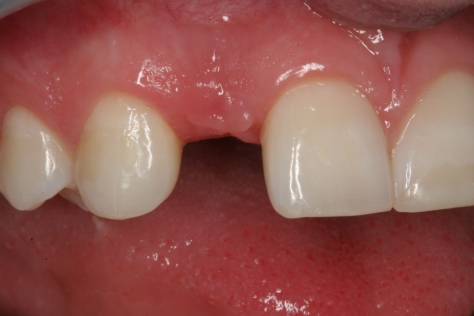
Edentulous space :
- The width should be enough for the implant (usually 5 to 7 mm).
- 1.5 mm to 2 mm of space is recommended between the head of the implant and the adjacent teeth.
- The traditional implant diameter is 3.75 mm (or 4 mm).
So the space of edentulous space should be 4 + 1.5 + 1.5 = 7 mm
- 1.5 mm is needed for the development and maintenance of the papilla.
Interradicular spacing :
- 5 mm is the minimum interradicular distance to place an implant.
- Orthodontist should align roots to make proper mesiodistal angulation of the roots in order to allow the surgeon to place the implant.
- Sometimes it is difficult even if the roots are made in better angulation.
In patients with class III tendency , as the crowns are tipped labially their roots will converge toward each other resulting in wagon wheel effect. The facial cortical plate limits the any labial root movement of maxillary incisors.
Papillary changes during space appropriation.:
When teeth are moved away from each other during space opening , the papilla remains stationary as the adjacent sulci are everted. The non-keratinized gingiva appears red but over time it keratinizes but location of the papilla does not change. This doesnt happen in a growing child.
Timing of implant placement :
- It is based on patient’s facial growth.
- By assessing growth by hand wrist x-rays or by evaluating serial cephalometric radiographs taken 6 months to 1 year apart.
Interim tooth replacement after orthodontics :
If implants cannot be placed until facial growth is complete , edentulous space is maintained by removable retainer with prosthetic tooth .
Implant placement :
- To ensure proper placement , a surgical guide should be fabricated.
- After placement 4 to 6 months should be allowed for adequate osseointegration to occur.
- A provisional restoration is placed before the final crown to allow the soft tissue to heal in its final position.
- The provisional restoration is allowed to remain in place for 4 to 6 weeks.
References :
- Kokich VG, Kinzer GA. Managing congenitally missing lateral incisors, part 1: Canine substitution. J
Esthet Restor Dent. 2005;17:1-6. - Kinzer GA, Kokich VO Jr. Managing congenitally missing lateral incisors. Part II: tooth-supported restorations. J
Esthet Restor Dent. 2005;17(2):76-84. - Kinzer GA, Kokich VO Jr. Managing congenitally missing lateral incisors. Part III: Single tooth-implant. J
Esthet Restor Dent. 2005;17:2-202-210.

Important topic, Thank you for your great efforts..
LikeLike
Thanks for encouraging and support.
LikeLike
Mashallah good job dr.Hani
LikeLike
Thanks for encouraging and support.
LikeLike