Category Archives: Diagnostic Procedures and Case Analysis

Early Transverse Treatment

Summarized and edited by : 
Dr. Amjad Alotaibi , Dr. Hani Alhebshi

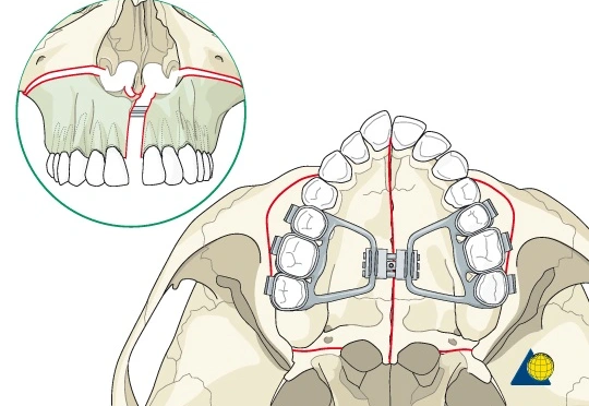
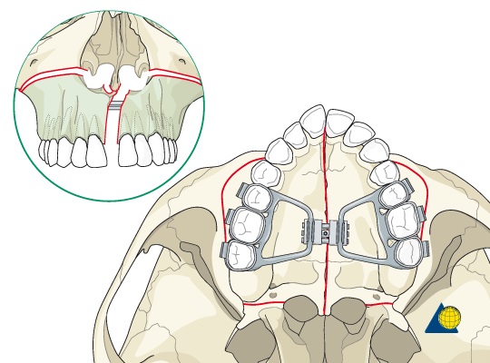
Expansion of the maxillary arch to improve transverse interarch relationships during the primary or mixed dentition stage is considered early transverse treatment as part of a two-phase treatment protocol. 

Three possible reasons for early transverse treatment:

  1. Correct posterior crossbite (The most common)
  2. Improve arch length deficiency 
  3. Facilitate correction of skeletal class II malocclusions 

early transverse tx copy copy

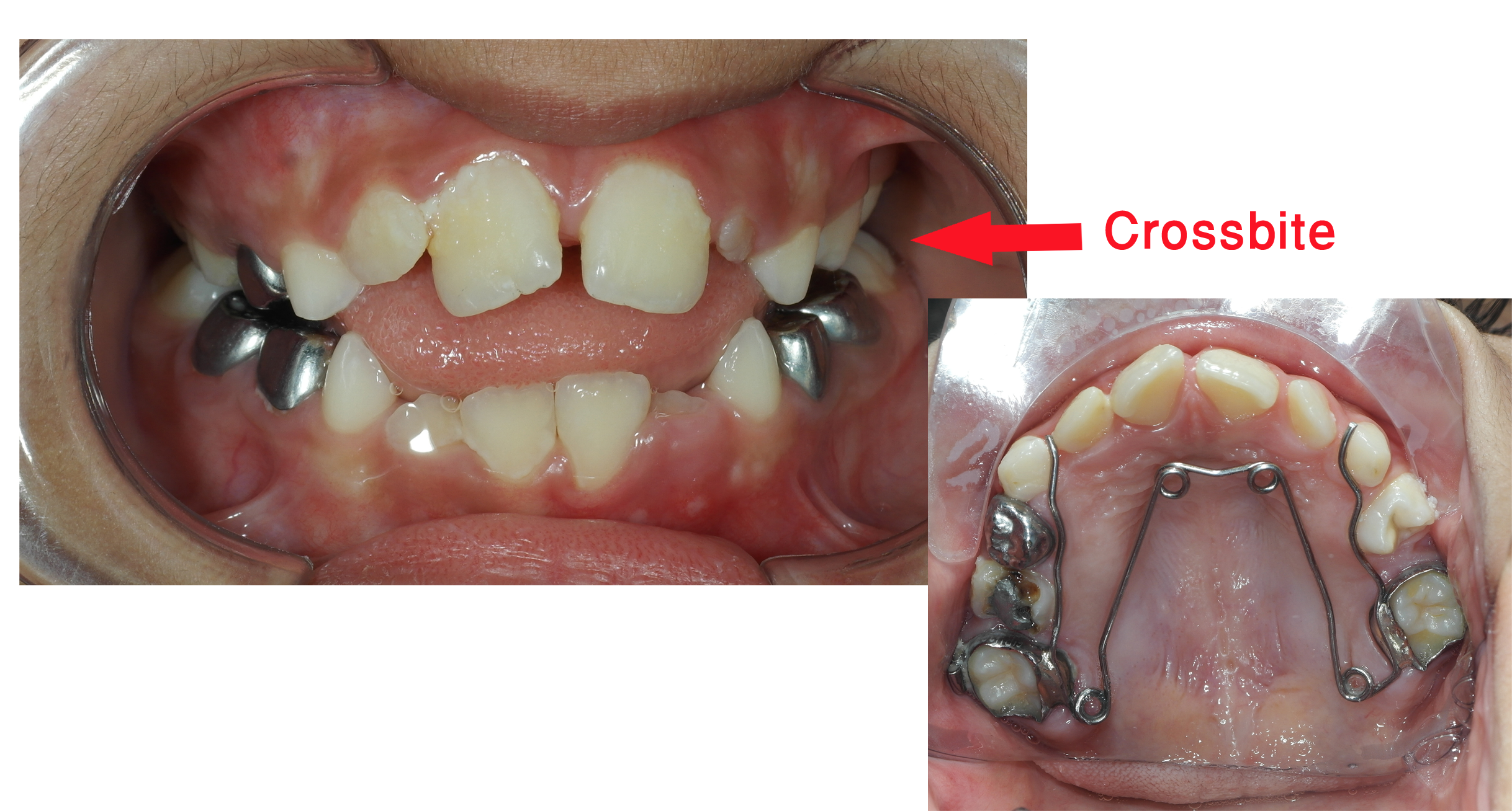
Posterior crossbite:

Types

Based on the position of the maxillary teeth:

  • Posterior lingual crossbite (most common)
  • Posterior buccal crossbite 

(N.B; Canine involvement is often seen and considered part of a posterior crossbite even though canines are not, by definition, posterior teeth) 

Based on the involved side of the arch :

  • Unilateral
  • Bilateral 

Incidence

In the primary and mixed dentition: Estimates range from 7% to 23% with a greater prevalence of unilateral crossbite coupled with a lateral shift of the mandible. 

Etiologies

A range of possible causes includes genetics, environmental factors, and habits that result in transverse maxillary skeletal deficiency, asymmetric growth of the maxilla or mandible, discrepant widths of basilar maxilla and mandible, and so on. 

Continue reading Early Transverse Treatment

Surgically Assisted Rapid Palatal Expansion

Surgically Assisted Rapid Palatal Expansion

Summarized by : Dr. Hani Alhebshi

Correction of Maxillary transverse deficiency (MTD) in a skeletally mature patient is more challenging than young patients because of changes in the osseous articulations of the maxilla with the adjoining bones. Surgically assisted rapid palatal expansion (SARPE) has gradually gained popularity as a treatment option to correct MTD. It allows clinicians to achieve effective maxillary expansion in a skeletally mature patient. The use of SARPE to treat MTD decreases unwanted effects of orthopedic or orthodontic expansion.

SARPE

Image source ( http://jawsurgeryforums.com/index.php?topic=6415.15 ).

Procedures to surgically correct MTD have conventionally been grouped into 2 categories: 

  • Segmenting the maxilla during a LeFort osteotomy to reposition the individual segments in a widened transverse dimension.
  • Surgically assisted rapid palatal expansion (SARPE). 


The criteria for selection of either of these to correct the MTD have not been clearly defined. The  reference of the surgeon often determines the choice of the procedure.

INDICATIONS FOR SARPE

There is a lack of consensus among orthodontists and surgeons about the indications for SARPE. 


The following have been reported in the literature as indications for SARPE, all applying to a skeletally mature patient with a constricted maxillary arch.


  1. To increase maxillary arch perimeter, to correct posterior crossbite, and when no additional surgical jaw movements are planned.
  2. To widen the maxillary arch as a preliminary procedure, even if further orthognathic surgery is planned. This is to avoid increased risks, inaccuracy, and instability associated with segmental maxillary osteotomy.
  3. To provide space for a crowded maxillary dentition when extractions are not indicated.
  4. To widen maxillary hypoplasia associated with clefts of the palate.
  5. To reduce wide black buccal corridors when smiling.
  6. To overcome the resistance of the sutures when OME has failed.


Diagnosis

There is much literature on the various methods used to diagnose MTD. 

  1. Clinical evaluation.
  2. Model analysis.
  3. Occlusograms.
  4. Radiographic measurements.

  Continue reading Surgically Assisted Rapid Palatal Expansion

Dental Traumas and their Orthodontic management

Summarized by : Dr. Hani Alhebshi

An orthodontist can face cases of dental trauma at his\her practice from time to time. These traumatic injuries differ in their types, severity and management.

This is a summary of the most common  situations of dental traumas and their management. This is based on two fantastic articles I strongly encourage to read.

Assessment and diagnosis of dental trauma

a comprehensive dental history which includes : asking about previous episodes of dental trauma or  treatment. 

a comprehensive clinical and radiographic assessment is essential prior to commencing orthodontic treatment and should include:


1. Hard tissue assessment

 a. Crown color assessment:

    • Dark hue might indicate loss of pulp vitality.
    • Yellow colour might indicate pulp canal obliteration
    • Pink colour might indicate internal resorption.

 b. Transillumination assessment can reveal enamel infraction lines and colour changes in traumatised teeth.


 c. Assessment of tooth mobility in both horizontal and vertical directions.


d. Percussion tests:

  • Tenderness to touching or tapping a tooth is suggestive of PDL damage.
  • High metallic percussion note is often diagnostic of ankylosis.
  • Dull percussion note may be suggestive of a root fracture.

 2. Soft tissue assessment:

a. Sinus tracts or swellings indicate peri-radicular infection.

b. Palpation over the tooth apex for tenderness as it might indicate a periapical infection.

 3. Radiographic assessment:

    • Two radiographs taken at different angulations are usually required to detect a root fracture. 
    • Radiographs can detect any root resorption or pulp canal obliteration due to previous trauma.
    • It is often very helpful if periapical radiographs of traumatized central incisors include both the injured tooth and its uninjured counterpart for direct comparison.

Continue reading Dental Traumas and their Orthodontic management

Management of congenitally missing lateral incisors

Summarized by : Dr Hani Alhebshi .

Three treatment options for replacing missing lateral incisors :

1.Canine substitution. ( Space Closure ).

2.A tooth supported restoration ( Space Opening ).

3.A single tooth implant ( Space Opening ).


The primary consideration to choose one option is to choose the most conservative one or the least invasive according to each case. 

congenitally missing lateral

Canine substitution :

Select the appropriate patient for canine substitution according to several criteria :

A. Malocclusion :

Two types of malocclusion can accept canine substitution according to
Kokich:

  1. Class II with no crowding in mandibular arch.In this pattern molars remain in class II and premolars are located in traditional canine position.
  2. Class I with crowding in lower arch that necessitate extraction.

Diagnostic wax up will greatly help the orthodontist and the dentist to evaluate the final occlusion and how much canine reduction is necessary.

B.Profile :

  • The ideal profile to accept canine substitution is straight profile or mildly convex. 
  • Moderately convex or retrusive mandible or chin are not appropriate patients.

Continue reading Management of congenitally missing lateral incisors