Introduction

بسم الله الرحمن الرحيم والصلاة والسلام على أشرف الأنبياء والمرسلين سيدنا محمد وعلى آله وصحبه أجمعين.

In the name of Allah, the most compassionate the most merciful.

G
In the beginning, I would like to welcome all visitors of our specialized blog.This blog was originally created to post , discuss and share scientific issues in Orthodontics especially what is related to periodic meetings held in King Abdulaziz University in Jeddah (Saudi Arabia ) as a part of the 4 to 5 years Saudi Board of Orthodontics program.

Thereafter ,the idea rose to another level to have many good articles of different orthodontic subjects summarized and posted here often by me and by some of our colleagues occasionally.These summaries I believe they would bring our clinical experience to a higher level in terms of clinical excellence.

I really encourage my orthodontic residents and specialists to participate by sharing their own useful summaries. You can share it here by sending it to our blog email (SorthoClub@gmail.com) and we will be so happy to added it here after reviewing the content.

Please ENJOY Orthodontics .. It is a wonderful specialty 🙂

Dr. Hani Alhebshi
Consultant Orthodontist : )

Surgical methods to uncover maxillary impacted canines

The following is a summary of an article done by Kokich that had shown the different techniques to uncover the maxillary impacted canines whether it was labially or palatally impacted.

Labial Impaction :

3 Techniques to uncover labial impactions  :

  • Excisional Uncovering.
  • Apical Repositioning Flap.
  • Closed Eruption technique.

Evaluate 4 criteria in labially impacted canines :

  1. Assess labiolingual position of canine :

    • If it is labially positioned use any of the 3 techniques because little bone pesent labially.
    • If it is in the center of the alveolous use Closed Eruption technique.
    • Other methods are difficult to perform because extensive bone might need to be removed occlusally.
  2. Assess vertical position relative to mucogingival junction.

    • Apical to Mucoginigival junction :
      • Deep to MGJ      :   Closed Eruption
      • Just above MGJ  : Apically repositioned flap mainly / Closed technique can be used too.
    • Coronal to Mucogingival Junction use Any of the 3 techniques.

Continue reading Surgical methods to uncover maxillary impacted canines

Endodontics – Orthodontics relation, progress and inter-action

Reviewed by : Dr. Moayyad Alkayyal

To look at the big picture, this review will focus on answering the following questions:

1.Is there an effect on the pulp during tooth movement?

2.Is it possible to move pulp-less teeth?

3.Is it possible to move actively inflamed teeth?

4.Is it safe to move previously traumatized teeth with pulpal involvement? What type of trauma? What type of movement?

5.Is there an increased risk of root resorption with RCT?

Continue reading Endodontics – Orthodontics relation, progress and inter-action

Canine Impactions

Topic prepared by Dr. Salwa Almadani …


INTRODUCTION

  • Tooth impaction can be defined as the infraosseous position of the tooth after the expected time of eruption.
  • Anomalous infraosseous position of the canine before the expected time of eruption can be defined as a displacement.
  • Early diagnosis and intervention could save the time, expense, and more complex treatment in the permanent dentition.

INCIDENCE

According to Thilander and  Ericson and Kurol :

The incidence of impacted maxillary canines is around   2-3% of the general population and mandibular canine impactions in approximately 0.2%.

Ngan P, Bishara the most common impacted tooth:

  • maxillary & mandibular 3rd Molars
  •   maxillary  canine
  •  mandibular  Premolars
  •  mandibular canine

Proffit, Cooke J:

  •  maxillary impaction  occur more commonly in females than males  in the ratio of 2:1

Stivaros and Mandall (2000)*

  •  Impacted canine crown is palatal in 61% of cases
  •  In the line of the arch in 34% of cases
  •  Labial in 4.5% of cases.


Prof.Afify AR,  Prof.Zawawi KH.  (2012)**

The prevalence of  impacted canine was the  second frequent anomaly  (3.3%) after congenitally missing teeth in the western region of Saudi Arabia. Continue reading Canine Impactions

General Guidelines to Management of Class II Malocclusion

Prepared by : Dr. Hani Alhebshi. , Dr, Saleh Alkhathami and Dr.Shehab Kaaki.

This is  a thorough overview on the management of  Calss II malocclusion.

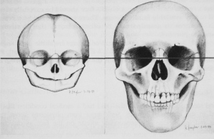
Signs of Class II malocclusion :

  • Convex Profile
  • Increased overjet
  • Skeletal pattern can be due to retrognathic mandible or prognathic maxilla or combination or both.That can be figured out by checking SNA and SNB angles.
  • Lips are commonly incompetent.
  • Nasiolabial angle is acute espaecially in Class II division I.
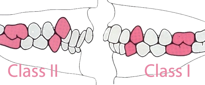
  • Class II molars , Canines and Incisors.
    British school classify malocclusions according to the incisors relation mainly , while american school mainly depends on the molar and canine relation.
    Therefore the British consider an increased overjet due to thumb sucking as Class II even if molars and canines are in Class I relation.

In Class II division 1 :

Treatment of class  II division  I is done sometimes to prevent trauma to maxillary anterior teeth because they are too proclined.

In Class II division 2 :
Upper central incisors are retroclined while laterals are in average or proclined inclination.
Overbite is commonly increased. The retroclined incisors might mask an increased overjet.Canine classification might then be a good parameter to check to confirm the Class II occlusion.

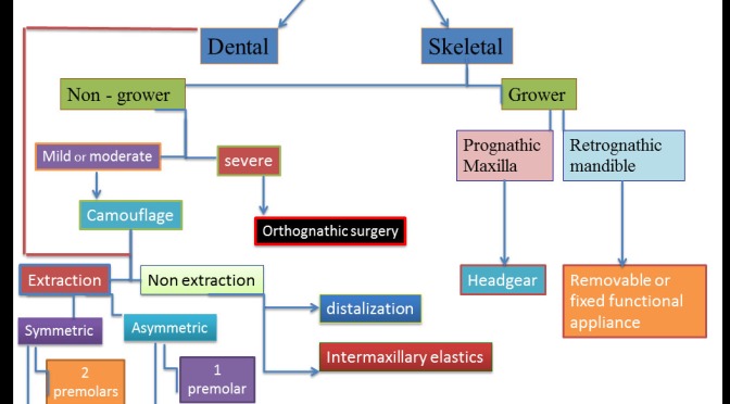
First step in managment : Determine the cause of Class II malocclusion. Is it skeletal or dental ?
If it is a skeletal then it can be due to a prognathic maxilla or a deficient mandible or combination of both.
On Cephalometric radiograph , ANB is oftenly increased ( above 4° ).

Skeletal Age : is the first important parameter that determines the path of treatment.In growing patients , growth modification can be used by the use of functional appliances or headgear while non-growing (adults) patients a camouflage or orthognathic surgery are chosen.

Growth Modification :
When patient is in preadolescence then a growth modification strategy can be considered.
CVM ( Cervical Vertebral Maturation) or hand wrist evaluation will show whether growth is still active or not.
Menarche ( The 1st menstruation cycle in girls )  indicates that growth has passed the peak in females. Ideal time to start growth modification is when CVM at stage 3 meaning that the peak in mandibular growth has occured within one year before this stage.
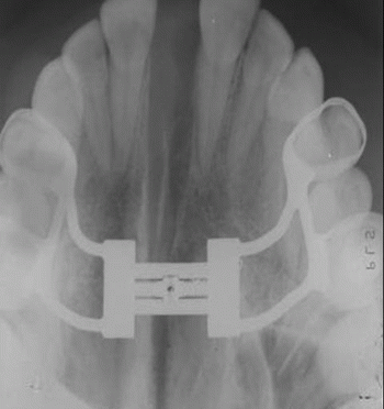
Therefore , correction of class II  can be carried out in this time by inducing mandibular advancement by functional appliances like herbst or twin block appliance. especially when the problem is mainly due to the mandibular retrognathism; or by restraining maxilla by means of headgear to allow mandible to grow and catch up maxillary growth especially when the problem is maxillary prognathism.
The choice between a high pull or cervical headgear depends on the vertical growth pattern of the patient.
If the lower facial height is high (with or without an open bite ) and mandibular plane angle is increased (hyperdivergent) then a high pull or combined headgear should be chosen to prevent extrusion of molars or any increase in the vertical dimension.
On the contrary, low facial height and (hypodivergent mandibular plane) necessate the use of cervical headgear type to allow some extrusion of molars and subsequent overbite correction owed to backward rotation of the mandible.

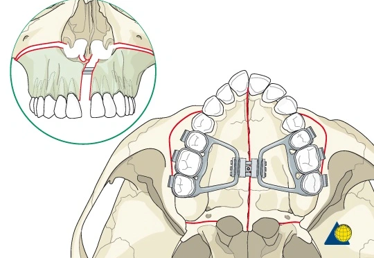
Orthognathic Surgery:

  • In late adolescence or adults (i.e non-growing): Usually a bilateral sagittal split osteotomy is performed to advance the mandible.
  • Maxillary setback with impaction can be beneficial too especially if the malocclusion is accompanied by a gummy smile.
  • Double jaw surgery can be done in both jaws if the problem is related to a combination of prognathic maxilla and retrognathic mandible.

The decision is better made by the surgeon and the orthodontist after making a visual treatment objective plan (VTO). This will predict the required movements needed to correct the skeletal discrepancy.
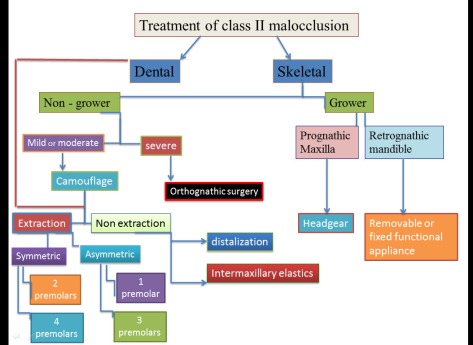
guidelines-tx-of-class-ii

Camuoflage :
Camouflage options are useful if patient has mild to moderate skeletal discrepancy or refuses surgical option.That will depend on the severity of Class II.
The soft tissue profile is a critical factor in the treatment plan.Nose and lips position are also other important parameters.

  • Camouflage treatment is most often done by extraction of upper 4s with maximum or absolute anchorage finishing molars in Class II while correcting canines into Class I relation.
  • Other options include extraction of upper 4s and lower 5s if crowding is severe in lower arch.
  • In severely flat profile or very prominent nose , extraction of upper premolars will ruin the profile and dish the face so that it appears uglier than before the treatment. Therefore , surgical approach should be presented to the patient as best option.
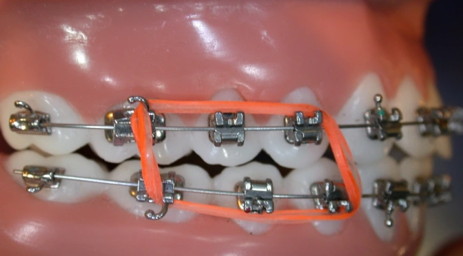
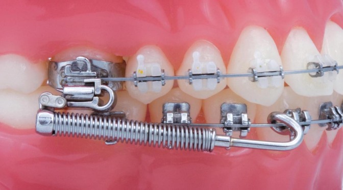
  • Using Class II correctors like Forsus is a good option unless patient has a severely proclined lower anterior teeth where it is contraindicated.
  • Class II elastics are also used to correct Class II molars and canines to class I relationship.It should be used cautiously especially when lower anteriors are proclined or vertical dimension is already high before treatment. Thats owing to the effect of class II elastics namely the protrusion of lower incisors , extrusion of lower molars that would increase the vertical dimension.Class II elastics is also contraindicated in gummy smile cases because of their effect in rotating the occlusal plane in a clockwise direction ending up with an increased gingival display.
  • Genioplasty ( to advance the chin )  is needed sometimes to correct the retrusive chin usually accompanied with extraction of upper 4s. this option is offered to the patient after the treatment of orthodontics is completed.

Effect of types of treatment on the soft tissue :

  • Lips should go back and become competent.
  • Nasiolabial angle should increase. ( i.e change from acute to obtuse angle. )


Orthognathic Surgery : How to

Edited by : Dr. Hani Alhebshi

Orthognathic surgery is a type of surgery that is done to the jaws to correct a frank skeletal discrepancy.

The common steps in order are :

  • Pre-surgical orthodontic treatment (decompensation orthodontics).  This involves aligning the teeth on the jaws according to their basal bone , and may require extractions,  A few weeks before surgery when decompensation orthodontics is completed patient will undergo a surgical assessment and pre-planning.  This involves taking impressions, x-rays, pictures and models of the teeth as well as a face bow (jaw registration).  This will allow the surgeon to plan the movements of the jaw and the technician will be able to construct the splints required during surgery.
  • The surgery is usually performed under general anaesthetic and can take up to 3 or 5 hours to complete depending on how much jaw surgery is required.   The new position of the jaws is maintained with the use of plates and screws and on occasions these may be resorbable .  Most common surgical cuts involve : Maxillary LeFort I for maxillary impaction or advancement , Bilateral sagittal split osteotomy for mandibualr advancement or set back , and  genioplasty to either advance or set back the chin.
  • Post-surgical Orthodontic Treatment (following surgery) : Elastics are applied to the teeth to guide the new bite and the patient is put on a soft diet for 6 weeks.  During this time medications such as antibiotics, analgesics and nasal drops are often required in the first week or two after surgery.  Then elastics are removed after about six weeks and the final adjustments to teeth and bite are undertaken by the orthodontist in consultation with the surgeon.  The process can last a further three to six months depending on the complexity of the procedure.

Here are videos that shows the procedures done in different types of orthognathic surgery.

Lefort 1 Osteotomy HD

Bilateral Sagittal Split osteotomy

Growth Mechanisms and regulation theories : A summary

Summarized by Dr.Hani Alhebshi ..

The aim of this summary is to outline the basic mechanisms and regulation theories  to be as a quick reference of craniofacial growth and development. More details can be found in the bottom of this summary when references are listed.


Craniofacial Mechanisms are devided into 4 components :

  • Growth Mechanisms ( How bone is formed ).
  • Growth Pattern ( change of size and shape of bone).
  • Growth Rate ( Speed at which bone is formed).
  • Regulation Mechanisms that regulate those three factors.


Growth Mehanisms :

Ossification of bone occurs in two ways :

  • Endochondral Ossification : Cartilage transformed into bone.
  • Intramembranous Ossification : Mesenchymal connective tissue is transformed into bone by deposition of bone on existing bone surfaces.

Continue reading Growth Mechanisms and regulation theories : A summary

Types of Arch wires

Summarized by Dr. Hani Alhebshi , from a course given by (Dr. Nabeel Talic from King Saud University). 

Classification of wires can be according to :

Material :

Stainless steel , Elgiloy , Titanium alloys , glass or polymers.

Coating :

Coated or non Coated : Ion implementation , Spray Coating , Sleeving.

Morphology :

Round , Square , Rectangular , Single or multi-strand.

Metal Wires :

  • Stainless steel
  • Cobalt Chromium ( Blue Elgiloy ).
  • B-Titanium Molybdenum ( TMA)
  • Titanium Niobium ( TiNb)
  • Nickle-Titanium ( NiTi).

Esthetic Wires

  • Plastic Coated wires
  • Composite wires.

Continue reading Types of Arch wires

When does midpalatal suture close ?

Summerized by : Dr.Hani Alhebshi.

When does midpalatal suture close ?

Upon searching the web , I found some useful articles answering this question.

It has been stated that there is quite wide variation among individuals that it ossifies at different age groups.

The amount of ossification increases in the growth spurt ( approximately 12-13 years in females , 14-15 years in males) leading to some obliteration at the suture.

There is evidence of continuous ossification in later ages upto 20s (young adults) and even to later age in other studies reaching 32 years! showing marked degree of closure.

The importance of this point lies in whether we can use rapid palatal expansion in adults or not to achieve an opening in the midpalatal suture.Several papers showed clinical cases that palatal sutures were opened in late teens and early twenties.

The following links are of valuable articles I found useful for our discussion.

Here are some important excerpts from these articles.

Continue reading When does midpalatal suture close ?

Nonsurgical RME (Haas) in Adults , is it reliable?

This post is prepared in collaboration with Dr. Maali Faqeeh ..

The results of the following papers indicate that nonsurgical RME in adults is a clinically successful and safe method for correcting transverse maxillary arch deficiency though it was thought to be an unreliable procedure.

1. Nonsurgical Rapid Maxillary Expansion in Adults: Report on 47 Cases Using the Haas Expander.

2. Nonsurgical Rapid Maxillary Alveolar Expansion in Adults: A clinical evaluation.

General Guidelines for Orthodontic Retention Protocol

Written By: Dr Rania AlRaddadi ..

As a continuation of the discussion we had today, I looked up some papers related to retention and it appears that no solid evidence is available up to date. The reason I believe is ofcourse the lack of well conducted trials and the huge variability in selecting the protocols among orthodontists.

Continue reading General Guidelines for Orthodontic Retention Protocol

Does Expansion add a value in absence of crossbite?

Written by : Dr. Waleed Farran ..

The recent trend in orthodontic diagnosis and treatment planning is toward an increasing emphasis on the soft tissue relationship and narrow buccal corridors, accompanied by a declining emphasis on the correction of malocclusion.However, depending of the upper arch as the base diagnostic arch can affect the stability of the orthodontic treatment.

This article discuss if there is no crossbite, did the expansion add a value…..?

Click here to download the article.


Surgery First Approach

Written by : Dr. Rania Alraddadi ..

Surgery First Approach

Dear all,

I wanted to share this article discussing this approach which is a systematic review.

In summary:

As we are all aware of its limitations and of the importance of case selection, the two major advantages for this approach are:

1- The immediate facial and esthetic improvement which is reflected on the patient’s psychological status.

2- The accelerated postoperative tooth movement as explained by the “RAP” phenomenon.

I also wanted to add some selection criteria for this approach which were mentioned by the maxillofacial team in our school:

1- Class III

2- No crowding

3- Flat curve of Spee

Have a great weekend!

Click here to download the article !


Finishing Cases with Class III Molars

Comment by Dr. Rania AlRaddadi ..

I want to attach a paper of 3 case reports which discusses different scenarios which all finished with Class III molars.

I think that the paper is beneficial in terms of expanding our horizon when it comes to different treatment options, especially and as we all know that the introduction of extraction treatment by Tweed made Angle’s sagittal molar relationship less critical in orthodontics.

The paper will follow.

( PDF ) STRATEGIES TO FINISH ORTHODONTIC TREATMENT WITH A CLASS III MOLAR RELATIONSHIP:THREE PATIENT REPORTS

Download paper 1 here

UPDATE 29/11/2016 : A new article related to the same topic is shared by Dr. Khalid Zawawi and Dr.Ehdaa Elshawley . Thanks to them.

Download paper 2 here

Orthodontic Brain storming